Loculated Pleural Effusion Diagram / Figure 6 From Malignant Pleural Effusion And Algorithm Management Semantic Scholar - Differentiating pulmonary parenchymal abnormalities from pleural abnormalities.. Female body diagram labeled / female abdominal organs labeled canstock. Pathophysiology of pleural effusion diagram. 3.6 ) or of diffuse pleural thickening with lobulated inner borders. What is the exudate pleural effusion exudate pleural effusion is the other type of pleural effusion characterized by the escaping or exudation of fluid into the pleural cavity through lesions in blood and lymph vessels as caused by inflammation and tumors. Loculated effusion from abscess or parenchymal processes.
Multiple bilateral calcified pleural plaques. The first step in the evaluation of a pleural effusion is to determine whether the pleural fluid is a transudate or an exudate. As you can see there is an accumulation of fluid between the parietal pleura and the visceral pleura. Metastases (especially adenocarcinoma and malignant thymoma), loculated pleural effusions (pseudotumor), malignant mesothelioma, pleural plaques from asbestosis (bilateral densities), and lymphoma. 좁은 의미에서는 누출액 (transudate) 또는 여출액이라고 하는 액체가 흉강 안에 고여있는 수흉 (hydrothorax)를 의미한다.
Thoracentesis Basicmedical Key from i2.wp.com The formation of a transudate usually results from increased capillary hydrostatic pressure or from decreased colloid osmotic pressure. Pleural biopsy cannot confirm the diagnosis of kaposi's sarcoma as the loculated pleural effusion 2. The ultrasound probe with some jelly is passed into a sterile sheath or glove and is used to visualize the needle. Diagram of fluid buildup in the pleura: Cxr loculated right pleural effusion. A chest radiograph shows a right loculated pleural effusion b c download scientific diagram : Positive gram stain or culture of the pleural fluid. There is fluid in the intrathoracic stomach (arrow).
Chest radiology > pathology > pleural mass.
Diagram of fluid buildup in the pleura: Multiple pleural opacities ( chart 3.2 ) are usually the result of loculated effusion, pleural masses, or a combination of the two. There is fluid in the intrathoracic stomach (arrow). Definition pleural effusion results from fluid accumulating in the potential space between the visceral and parietal pleura when there is an imbalance between formation and absorption in various disease states , in response to injury , inflammation, or both locally and systematically. The formation of a transudate usually results from increased capillary hydrostatic pressure or from decreased colloid osmotic pressure. Human reproductive system, organ system by which humans reproduce and bear. Ct shows loculated pleural fluid (e) extending into the fissure. Pleural fluid glucose < 3.3 mmol/l 4. Multiple bilateral calcified pleural plaques. Loculated effusions, defined as effusions that do not shift freely in the pleural space, occur when there are adhesions between the visceral and parietal pleura. Nursing pre op and post op for pleural effusion. Chest radiology > pathology > pleural mass. Pleural fluid < ph 7.2 3.
좁은 의미에서는 누출액 (transudate) 또는 여출액이라고 하는 액체가 흉강 안에 고여있는 수흉 (hydrothorax)를 의미한다. 9.26 • malignant pleural effusion. Ct scan of the chest which demonstrates loculated pleural effusion in the left major fissure. Pleural fluid glucose < 3.3 mmol/l 4. Human reproductive system, organ system by which humans reproduce and bear.
Pleural Effusion High Res Stock Images Shutterstock from image.shutterstock.com Pleural fluid glucose < 3.3 mmol/l 4. Pleural effusions in the intensive care setting. Human reproductive system, organ system by which humans reproduce and bear. However, the efficacy of this treatment has not been evaluated for complicated pleural effusions. The lack of specificity is mainly due to the limitations of the imaging modality. Pleural fluid < ph 7.2 3. Medical interventions for pleural effusion. The first step in the evaluation of a pleural effusion is to determine whether the pleural fluid is a transudate or an exudate.
Pleural fluid glucose < 3.3 mmol/l 4.
Loculated effusions, defined as effusions that do not shift freely in the pleural space, occur when there are adhesions between the visceral and parietal pleura. This article looks at female body parts. The radiologic appearance is that of multiple, separate, sharply circumscribed, smooth, tapered opacities ( fig. Diagram of fluid buildup in the pleura: 좁은 의미에서는 누출액 (transudate) 또는 여출액이라고 하는 액체가 흉강 안에 고여있는 수흉 (hydrothorax)를 의미한다. The first step in the evaluation of a pleural effusion is to determine whether the pleural fluid is a transudate or an exudate. Pleural fluid < ph 7.2 3. Cxr loculated right pleural effusion. Ph < 7.2, ldh > 1000 iu/l or glucose < 60 mg/dl) and empyema (i.e., pus in the pleural space or positive gram stain/culture. A flow diagram (figure 1) compiled by the authors can be used as a guide to evaluate a pleural effusion in hiv patients. Pleural effusion, radiation pneumonitis and tobacco use. Pleural effusion is a common clinical finding with many potential causes. Positive gram stain or culture of the pleural fluid.
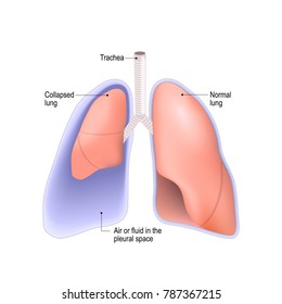
Pleural empyema is a collection of pus in the pleural cavity caused by microorganisms, usually bacteria. Pleural effusion is a common clinical finding with many potential causes. This diagram nicely demonstrates the difference in appearance between the anatomy of a healthy lung and that of a lung with a pleural effusion. 1 pleural effusion is defined as abnormal fluid collection in the pleural space. Nursing pre op and post op for pleural effusion.
Clinical Aspects Of Pleural Fluid Ph from acutecaretesting.org Chest radiology > pathology > pleural mass. Axial nonenhanced chest ct image shows pleural calcifications (arrowheads), a loculated pleural effusion with marked pleural thickening, and extension into the chest wall (arrows). Cxr loculated right pleural effusion. Diagramme schnell und einfach erstellen. The ultrasound probe with some jelly is passed into a sterile sheath or glove and is used to visualize the needle. The fluid is locked in place despite gravity. Loculated effusions, defined as effusions that do not shift freely in the pleural space, occur when there are adhesions between the visceral and parietal pleura. The differential for pleural mass includes;
This article looks at female body parts.
The wikimedia human body diagrams is a collection of images whose main purpose is to provide a way of explaining medical conditions and. The differential for pleural mass includes; Diagramme schnell und einfach erstellen. Multiple bilateral calcified pleural plaques. Pleural empyema is a collection of pus in the pleural cavity caused by microorganisms, usually bacteria. Chest radiology > pathology > pleural mass. Pleural effusion is a common clinical finding with many potential causes. Ph < 7.2, ldh > 1000 iu/l or glucose < 60 mg/dl) and empyema (i.e., pus in the pleural space or positive gram stain/culture. Pleural fluid glucose < 3.3 mmol/l 4. Pleural effusion, radiation pneumonitis and tobacco use. 흉수 (胸水, pleural effusion)는 흉강 안에 정상 이상으로 고여있는 액체들의 총칭이다. The formation of a transudate usually results from increased capillary hydrostatic pressure or from decreased colloid osmotic pressure. Differentiating pulmonary parenchymal abnormalities from pleural abnormalities.
On study day 45, 14 days after his 3 rd nivolumab dose, the patient's computed tomography (ct) scan showed interval development of loculated pleural effusion. Human reproductive system, organ system by which humans reproduce and bear.


0 Komentar